Filament tracing

Filament tracing operations are image analysis operations in which there is an image of a filamentous structure (it may be a tree-like structure, a filament network or a agglomeration of single 'stick-like' filaments) as input and outputs data that represent the filament, most commonly a skeleton representation of the filaments and their diameters or surfaces.

Synonyms
Tubular structure extraction
biofilament tracing
Curvilinear structure reconstruction
Curvilinear structure detection
neuron image analysis
neuron reconstruction
Description

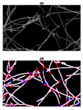
Trace ridges combines filtering, Watershed transforms, Edge detection and mathematical morphology to trace ridges in an image with fibre-like structures. 
Publication provides objective comparison of six existing methodologies (Edge detection, CT Fire, Scale Space, Twombli, U-Net and Graph Based).

has function
Description

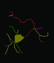
An imageJ/Fiji plugin that measures and classifies neurites from a very large number of neurons.

SNT

Description

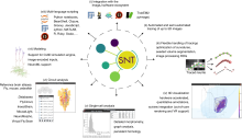
SNT is ImageJ’s framework for tracing, visualization, quantitative analyses and modeling of neuronal morphology. For tracing, SNT supports modern multidimensional microscopy data, semi-automated and automated routines, and options for editing traces. For data analysis, SNT features advanced visualization tools, access to all major morphology databases, and support for whole-brain circuitry data.

Schematic Overview of SNT components and SNT functionality
Description

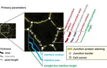
Junction Mapper is a semi-automated software (Java Desktop application) for analysing data from images of cells in close proximity to each other in monolayers. The focus of Junction Mapper is to measure the morphology of cell boundaries, define single junctions and quantify the length, area and intensity of the staining of different proteins localised at cell-cell contacts. The output are various unique parameters that assess the contacting interface between cells and up to two junctional markers.

junction mapper
Description

ClearMap is a toolbox for the analysis and registration of volumetric data from cleared tissues.

It was initially developed to map brain activity at cellular resolution in whole mouse brains using immediate early gene expression. It has since then been extended as a tool for the qunatification of whole mouse brain vascualtur networks at capilary resolution.

It is composed of sevral specialized modules or scripts: tubemap, cellmap, WobblyStitcher.

ClearMap has been designed to analyze O(TB) 3d datasets obtained via light sheet microscopy from iDISCO+ cleared tissue samples immunolabeled for proteins. The ClearMap tools may also be useful for data obtained with other types of microscopes, types of markers, clearing techniques, as well as other species, organs, or samples.

ClearMap SCreenshot