web

Description

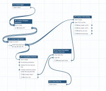
This workflow is the integration of YOLO (You Only Look Once) machine learning models, image pre-processing scripts and labeling tools within the Galaxy platform. Galaxy is an open, web-based platform used primarily for data analysis in computational biology, but it also has applications in image processing and other fields. 

How the Galaxy YOLO image segmentation tool works

The combination of Galaxy and YOLO allows researchers to perform object detection and image analysis without requiring extensive programming knowledge. Here's how it generally works: 

  • Web-based interface: Galaxy provides a graphical, user-friendly interface to access powerful analysis tools. Users can simply upload their image data, select the YOLO tool, and run the analysis.
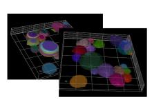
  • YOLO model execution: The Galaxy tool executes a pre-trained YOLO model, often from the Ultralytics framework, on the input images. These models can perform tasks like object detection (drawing bounding boxes) or instance segmentation (creating pixel-level masks).
  • Training and prediction: Some tools allow for both model training and prediction. Users can train a custom YOLO model on their own labeled datasets to detect specific objects of interest. For example, bioimage analysis may involve detecting cells or other structures.
  • Other integrations: Other machine-learning tools can be integrated with YOLO in Galaxy. For instance, the AnyLabeling tool supports YOLO for semi-automated and active learning-based data annotation. 
Description

BIIGLE is a web-based software for image and video annotation that enables collaborative research on large datasets. It offers tools for manual and computer-assisted annotation, quality control and the collaboration on custom taxonomies to describe objects. BIIGLE is freely available and can be installed in cloud environments, a local network or on mobile platforms during research expeditions. The public instance on biigle.de is free for non-commercial use.

BIIGLE Logo
Description

TissUUmaps is a browser-based tool for fast visualization and exploration of millions of data points overlaying a tissue sample. TissUUmaps can be used as a web service or locally in your computer, and allows users to share regions of interest and local statistics.

Description

While a quickly retrained cellpose network (only on xy slices, no need to train on xz or yz slices) is giving good results in 2D, the anisotropy of the SIM image prevents its usage in 3D. Here the workflow consists in applying 2D cellpose segmentation and then using the CellStich libraries to optimize the 3D labelling of objects from the 2D independant labels.

Here the provided notebook is fully compatible with Google Collab and can be run by uploading your own images to your gdrive. A model is provided to be replaced by your own (create by CellPose 2.0)

has function
example of usage
Description

SuperDSM is a globally optimal segmentation method based on superadditivity and deformable shape models for cell nuclei in fluorescence microscopy images and beyond.